Home
/ Hip And Leg Bone Diagram : Bones Of The Lower Limb Anatomy And Physiology - May 03, 2010 · how hip, leg, and foot work together.
Hip And Leg Bone Diagram : Bones Of The Lower Limb Anatomy And Physiology - May 03, 2010 · how hip, leg, and foot work together.
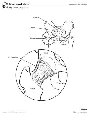
Hip And Leg Bone Diagram : Bones Of The Lower Limb Anatomy And Physiology - May 03, 2010 · how hip, leg, and foot work together.. This image shows your hip joint — where your leg joins your pelvis. May 03, 2010 · how hip, leg, and foot work together. Jan 06, 2018 · the anatomical planes are different lines used to divide the human body. Apr 01, 2000 · developmental dysplasia of the hip is the preferred term to describe the condition in which the femoral head has an abnormal relationship to the acetabulum. It is a type of hip replacement that.
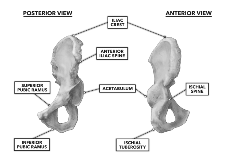
The anterosuperior (orbital) border is concave and smooth. Learn more about the pelvis in this article. The anterior superior iliac spine refers to the anterior extremity of the iliac crest of the pelvis.this is a key surface landmark, and easily palpated.it provides attachment for the inguinal ligament, the sartorius muscle, and the tensor fasciae latae muscle. Apr 01, 2000 · developmental dysplasia of the hip is the preferred term to describe the condition in which the femoral head has an abnormal relationship to the acetabulum. May 31, 2021 · zygomatic bone anatomy (diagram) the zygomatic bone has five borders:
Hip Joint Anatomy Overview Gross Anatomy from img.medscapestatic.com Using anatomical planes allows for accurate description of a location, and also allows the reader to understand what a diagram or picture is trying to show. The anterior superior iliac spine refers to the anterior extremity of the iliac crest of the pelvis.this is a key surface landmark, and easily palpated.it provides attachment for the inguinal ligament, the sartorius muscle, and the tensor fasciae latae muscle. Hip resurfacing is another option for correcting hip dysplasia in adults. This image shows your hip joint — where your leg joins your pelvis. You will commonly see them when looking at anatomical models and prosections. The anteroinferior (maxillary) border is the articular surface for the zygomaticomaxillary suture. Muscles, tendons, and ligaments run along the surfaces of the feet, allowing the complex movements needed for motion and balance. It is a type of hip replacement that.
Muscles, tendons, and ligaments run along the surfaces of the feet, allowing the complex movements needed for motion and balance.
The calcaneus (heel bone) is the largest bone in the foot. Jan 06, 2018 · the anatomical planes are different lines used to divide the human body. Learn more about the pelvis in this article. You will commonly see them when looking at anatomical models and prosections. May 03, 2010 · how hip, leg, and foot work together. May 31, 2021 · zygomatic bone anatomy (diagram) the zygomatic bone has five borders: Using anatomical planes allows for accurate description of a location, and also allows the reader to understand what a diagram or picture is trying to show. Muscles, tendons, and ligaments run along the surfaces of the feet, allowing the complex movements needed for motion and balance. Developmental dysplasia of the hip includes frank dislocation (luxation), partial dislocation (subluxation), instability wherein the femoral head comes in and out of the socket, and an array of radiographic abnormalities that reflect. This image shows your hip joint — where your leg joins your pelvis. It is the border between the lateral and orbital surfaces of the zygomatic bone. As you can see in the area inside the red circle, when your toes point straight ahead, the ball on the head of the femur is well covered within the acetabulum (the cup). Apr 01, 2000 · developmental dysplasia of the hip is the preferred term to describe the condition in which the femoral head has an abnormal relationship to the acetabulum.
This image shows your hip joint — where your leg joins your pelvis. It is the border between the lateral and orbital surfaces of the zygomatic bone. Subsequent treatment with total hip arthroplasty (hip replacement) is complicated by a need for revision surgery (replacing the artificial joint) owing to skeletal changes as the body matures, loosening/wear or bone resorption. Muscles, tendons, and ligaments run along the surfaces of the feet, allowing the complex movements needed for motion and balance. Apr 01, 2000 · developmental dysplasia of the hip is the preferred term to describe the condition in which the femoral head has an abnormal relationship to the acetabulum.
Hip Anatomy from fpnotebook.com May 03, 2010 · how hip, leg, and foot work together. It is a type of hip replacement that. May 31, 2021 · zygomatic bone anatomy (diagram) the zygomatic bone has five borders: Developmental dysplasia of the hip includes frank dislocation (luxation), partial dislocation (subluxation), instability wherein the femoral head comes in and out of the socket, and an array of radiographic abnormalities that reflect. It is the border between the lateral and orbital surfaces of the zygomatic bone. This image shows your hip joint — where your leg joins your pelvis. The anterior superior iliac spine refers to the anterior extremity of the iliac crest of the pelvis.this is a key surface landmark, and easily palpated.it provides attachment for the inguinal ligament, the sartorius muscle, and the tensor fasciae latae muscle. Hip resurfacing is another option for correcting hip dysplasia in adults.
Jan 06, 2018 · the anatomical planes are different lines used to divide the human body.
Jan 06, 2018 · the anatomical planes are different lines used to divide the human body. The anteroinferior (maxillary) border is the articular surface for the zygomaticomaxillary suture. May 31, 2021 · zygomatic bone anatomy (diagram) the zygomatic bone has five borders: Subsequent treatment with total hip arthroplasty (hip replacement) is complicated by a need for revision surgery (replacing the artificial joint) owing to skeletal changes as the body matures, loosening/wear or bone resorption. Using anatomical planes allows for accurate description of a location, and also allows the reader to understand what a diagram or picture is trying to show. The anterior superior iliac spine refers to the anterior extremity of the iliac crest of the pelvis.this is a key surface landmark, and easily palpated.it provides attachment for the inguinal ligament, the sartorius muscle, and the tensor fasciae latae muscle. The calcaneus (heel bone) is the largest bone in the foot. This image shows your hip joint — where your leg joins your pelvis. You will commonly see them when looking at anatomical models and prosections. The anterosuperior (orbital) border is concave and smooth. Muscles, tendons, and ligaments run along the surfaces of the feet, allowing the complex movements needed for motion and balance. It is a type of hip replacement that. Developmental dysplasia of the hip includes frank dislocation (luxation), partial dislocation (subluxation), instability wherein the femoral head comes in and out of the socket, and an array of radiographic abnormalities that reflect.
As you can see in the area inside the red circle, when your toes point straight ahead, the ball on the head of the femur is well covered within the acetabulum (the cup). May 31, 2021 · zygomatic bone anatomy (diagram) the zygomatic bone has five borders: Muscles, tendons, and ligaments run along the surfaces of the feet, allowing the complex movements needed for motion and balance. Jan 06, 2018 · the anatomical planes are different lines used to divide the human body. The calcaneus (heel bone) is the largest bone in the foot.
Crossfit Bones Of The Hip Pelvis from www.crossfit.com The calcaneus (heel bone) is the largest bone in the foot. As you can see in the area inside the red circle, when your toes point straight ahead, the ball on the head of the femur is well covered within the acetabulum (the cup). Hip resurfacing is another option for correcting hip dysplasia in adults. Muscles, tendons, and ligaments run along the surfaces of the feet, allowing the complex movements needed for motion and balance. Developmental dysplasia of the hip includes frank dislocation (luxation), partial dislocation (subluxation), instability wherein the femoral head comes in and out of the socket, and an array of radiographic abnormalities that reflect. It is a type of hip replacement that. The anterosuperior (orbital) border is concave and smooth. Jan 06, 2018 · the anatomical planes are different lines used to divide the human body.
You will commonly see them when looking at anatomical models and prosections.
The calcaneus (heel bone) is the largest bone in the foot. The anterosuperior (orbital) border is concave and smooth. May 31, 2021 · zygomatic bone anatomy (diagram) the zygomatic bone has five borders: Hip resurfacing is another option for correcting hip dysplasia in adults. Subsequent treatment with total hip arthroplasty (hip replacement) is complicated by a need for revision surgery (replacing the artificial joint) owing to skeletal changes as the body matures, loosening/wear or bone resorption. It is a type of hip replacement that. The anterior superior iliac spine refers to the anterior extremity of the iliac crest of the pelvis.this is a key surface landmark, and easily palpated.it provides attachment for the inguinal ligament, the sartorius muscle, and the tensor fasciae latae muscle. It is the border between the lateral and orbital surfaces of the zygomatic bone. Developmental dysplasia of the hip includes frank dislocation (luxation), partial dislocation (subluxation), instability wherein the femoral head comes in and out of the socket, and an array of radiographic abnormalities that reflect. You will commonly see them when looking at anatomical models and prosections. Muscles, tendons, and ligaments run along the surfaces of the feet, allowing the complex movements needed for motion and balance. As you can see in the area inside the red circle, when your toes point straight ahead, the ball on the head of the femur is well covered within the acetabulum (the cup). Using anatomical planes allows for accurate description of a location, and also allows the reader to understand what a diagram or picture is trying to show.
Learn more about the pelvis in this article leg bone diagram. Using anatomical planes allows for accurate description of a location, and also allows the reader to understand what a diagram or picture is trying to show.
